Tendons are tough, fibrous tissues that attach muscles to bone. The Achilles tendon, located just above the heel of your foot, attaches the gastrocnemius muscle (Fig. 1A) and the soleus muscle (Fig. 1B) to the back of the heel. Another name for the muscle group around the Achilles tendon is “triceps surae.” The Achilles tendon transmits the muscular force of the triceps surae to the ankle and foot lever. Achilles tendonitis is a common overuse injury seen in people who do activities involving running or jumping; such repetitive motions can lead to tendon damage. Analyzing the dynamics between the triceps surae, the Achilles tendon, and the ankle/foot during movement will make it easier to understand why Achilles tendonitis occurs and how to treat and prevent it.
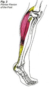
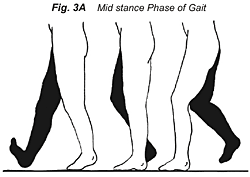
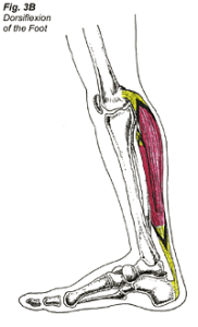
There are two types of muscle contractions: a concentric contraction, and an eccentric contraction. A concentric contraction of the triceps surae would shorten the length of the muscle, causing the Achilles tendon to pull on the back of the heel bone, resulting in the heel lifting up and the toes pointing downward. This particular motion is termed plantar flexion of the ankle/foot (Fig. 2). An eccentric contraction of the triceps surae would prevent injury by slowing down or limiting excess motion of that area. After the foot strikes the ground and enters the mid stance phase of gait (Fig. 3A), the combination of ground reactive forces on the foot and momentum from forward leg movement results in dorsiflexion of the foot (Fig. 3B). During the mid stance phase of gait, the triceps surae contracts, causing the Achilles tendon to resist this dorsiflexion of the foot. The mid stance phase of gait ends when the heel lifts off of the ground surface.
 Repetitive use of the Achilles tendon and triceps surae occurs during the midstance phase of gait/motion, and can result in trauma to the area. Tendons have fewer blood vessels than muscles, which means that less oxygen and nutrients can get to them. Faced with this shortage of blood supply, tendons take a longer time to heal than muscles. If the trauma to the tendon is faster than the rate of healing and repair, Achilles tendonitis will result.
There are five grades of tendonitis:
Grade 1 – Pain does not occur during activity, but generalized pain will be felt in the Achilles region after the training session has ended. Tenderness in the Achilles tendon will resolve before the next training session.
Grade 2 – Minimal localized pain will be present in the Achilles tendon during the training session; the pain will disappear before the next training session.
Grade 3 – Pain in the Achilles tendon now interferes with the training session but disappears before the next training session.
Grade 4 – Pain in the Achilles tendon severely interferes with the training sessions and does not disappear before the next training session.
Grade 5 – Pain in the Achilles tendon interferes with the training sessions and activities of daily living. The Achilles tendon becomes deformed and there is a loss of function of the triceps surae.
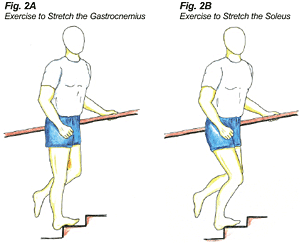
 Treatment at Dr. Dubin’s office of a grade 1 or a grade 2 tendonitis would consist of adjustments to the ankle and foot to free up joint motion, deep tissue to the musculature of the and leg to free up soft tissue motion, and ultrasound and electric muscle stimulation combotherapy to restore normal tone, help in the healing process, and decrease pain. The patient would be encouraged to ice the Achilles tendon for 20 minutes after each training session. A strength-training program for the lower extremities would be implemented, emphasizing eccentric strength training of the Achilles tendon. Stretching of the Achilles musculature and tendon could be conducted on a step (Fig. 4A & 4B), and a stretching routine for the lower extremities utilizing the Flexband® would be encouraged.
 Semi-rigid orthotics with no more than 7/8″ medial arch support can be a useful tool in the prevention of Achilles tendonitis in runners who have high arches or flat feet. A temporary 1/8″ heel lift can also be added to the orthotic to limit dorsiflexion of the foot; this would take pressure off of the injured Achilles tendon. Achilles tendonitis is common in recreational runners who act as their own coaches. These runners may prematurely increase the intensity, duration and frequency of their running sessions. Proper training techniques would allow for adaptive changes of the Achilles tendon, including strengthening of the tendon and increased blood supply to the area. These changes will increase the ability of the Achilles to accept increased activity but they take time and patience. For marathon runners, initially a training base of four miles needs to be established at 65%-75% maximum heart rate. Later, a training schedule should be followed. A proper warm-up consisting of a jog will increase the blood supply to the muscles and tendons, making them more efficient in absorbing loads. Hill training should gradually be added to a training route because uphill running will increase the eccentric load on the Achilles tendon. Runners should also change their every 250-400 miles, during which their sneakers have lost approximately 40% of their shock absorption capabilities.
Early therapy and intervention is important in the prevention of a grade 3 in progressing to a grade 4 or a grade 5 tendonitis. In the early stages of a grade 3 tendonitis, one week of rest from the offending activity should occur, as well as similar treatment procedures as administered in a grade 1 or a grade 2 tendonitis. Cross training with swimming, bicycling, or an elliptical machine would be useful in maintaining aerobic capacity and in allowing the Achilles tendon and triceps surae to heel properly before resumption of training.

Treatment of a more advanced grade 3 tendonitis or grade 4 tendonitis will involve a longer rest from the offending activity and a slower progression of weight training and stretching. Understanding the 3 phases of repair of injured tissues is necessary for proper treatment of these injuries.
The 3 phases of repair are the reactive phase, regenerative phase, and the remodeling phase. The reactive phase is the initial inflammatory response to trauma, where vasodilation of the blood vessels surrounding the injured tendon or muscle occurs, causing swelling, pain, and loss of function. To limit the immobilizing effects of the reactive phase, RICE (rest, ice, compression, and elevation) should be applied to the injured region. During the regenerative phase, dead cells are cleared out; tiny blood vessels are restructured to help supply oxygen to the damaged tissue; and collagen is laid down to help repair the injured tissue. Collagen, the connective tissue that replaces the damaged tendon or muscle, is initially weak, but with time, its strength improves.
After 7-14 days, damaged muscles regain approximately 50% of their strength; tendons may take a longer time to regain their strength. During the remodeling phase, the collagen tissue matures, and with proper medical treatment and compliance with a strength training and flexibility routine, the tissue will hopefully regain 100% strength. The remodeling phase can sometimes take up to six months for muscle repair and even longer for tendon repair, depending on the severity of the injury.

